University of Arizona researchers have been awarded a $2 million grant to perfect their bone-regrowth technology using a combination of 3D printing and adult stem cells that one day may help wounded soldiers.
The five-year grant, issued by the Department of Defense to UA professor of orthopedic surgery and biomedical engineer John Szivek and his team, will be used to study ways to quickly regenerate long segments of missing bone and how exercise affects the speed of recovery.
The DOD is particularly interested in developing the technology to treat wounded military personnel, but the technology would ultimately be useful for a variety of injuries.
Car crashes produce the most common injuries that would require this type of treatment, said David Margolis, co-investigator on the grant and doctor of orthopedic surgery. It can also be used to reconstruct bone removed in surgical cancer treatment, Szivek said.
Currently, a common solution for bone-shattering injuries is to replace the missing segment with a support rod and cadaver bone.
The living bone connects to the cadaver bone at each end, but doesn’t connect across it to create one long piece of living bone, which is necessary for healing.
“The DOD knows that this doesn’t work well,” Szivek said. The dead cadaver bone will begin to crack and eventually break. For active people, this method lasts only a couple of years.
Other options are also available, Margolis said, but all current options and outcomes can be frustrating for patients.
“Nothing is guaranteed to work and each one has different risks and benefits,” he said. “None give you back what you had before.”
Skeletal seeds
“We’re trying to find a way to regrow that segment of bone using a person’s own living cells,” Szivek said.
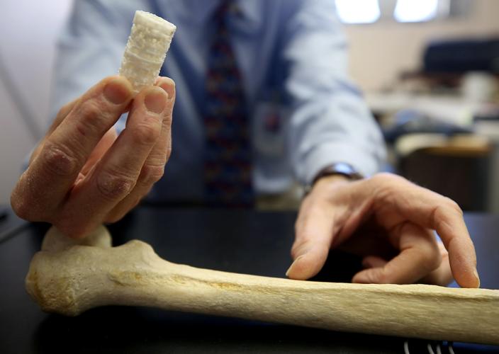
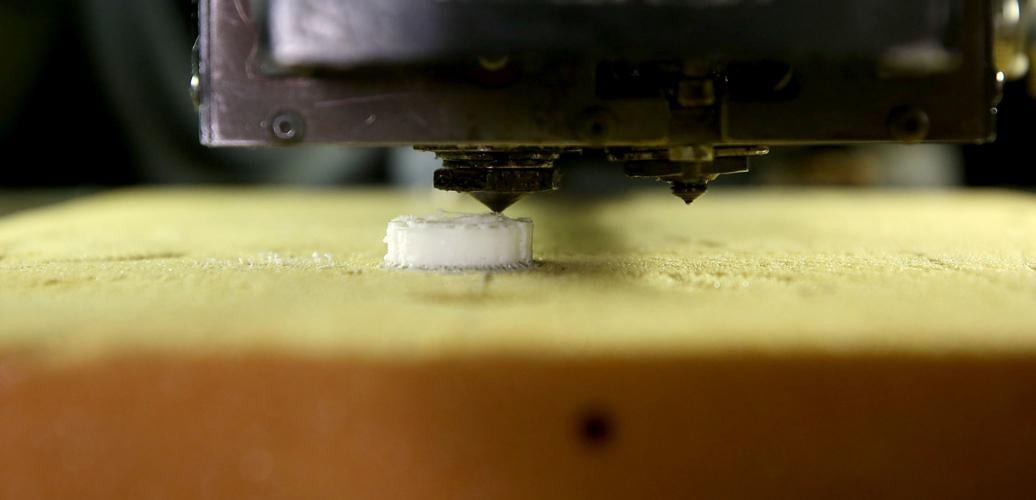
To do so, he and his team designed and 3D-printed a piece of plastic scaffolding hollowed out with grooves and small cavities. The final product has a structure similar to a sponge. The team chose the shape to mimic the natural structure of bone.
The 3D-printed scaffolding shape can be customized to the specific needs of the patient, Margolis said.
“We can even scan the other limb to get an idea and print the mirror image if we needed to.”
They will then seed the scaffold with the patient’s stem cells and calcium “to jump-start the process,” Szivek said. Adult stem cells are capable of evolving into bone cells that build bone tissue, and the calcium is an additional cue for the stem cells that pushes them to work faster.
Working smarter and harder
Before applying for the grant, the team did a pilot study on sheep to test its methods. They were all shocked by how quickly they were able to generate bone, but they want to push it even faster.
Szivek said they want to be able to bridge two separate pieces of bone as quickly as possible because “your body has a tendency to stop regeneration of bone tissues after about six months and instead starts building scar tissue.”
The pilot study showed complete bridging between bone segments within a month or two, “which is really fast,” Szivek said.
Complete bridging along three surfaces, which should be enough to support a person, was reached within about three months.
Within six months, the team began to see remodeling, which is the process of smoothing out the new bone and adapting to the demands placed on it.
For example, “If you suddenly decided to be an Olympic weightlifter, within months, your bones would get thicker because they’re adapting to what you need to do,” Szivek said. Conversely, the bones of inactive people are thinner.
“We really want to push the system to run as quickly as possible. We want it to form any kind of bone at all. It doesn’t matter, it could be the crappiest bone on earth, but once it’s there, it will remodel itself,” he said. “And once it does that, it will adapt to the exercise level of the person.”
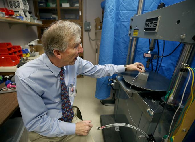
With the grant, Szivek also plans to embed sensors into the new bone to generate feedback on the effect exercise has on the remodeling and healing process.
“Physical therapists could use this information to do exercises with patients so they can heal effectively once we know the relationship between exercise and healing,” Szivek said.
Eventually, he envisions these implanted sensors sending updates to patients’ phones that will tell them they’re running too fast or they’re jumping too hard.
For now, he’s using animal models but thinks that by the end of the grant, he can test the technology on a limited number of human volunteers, he said.
This is important because with animal models, “You’re always using a healthy animal, but most people who have bad injuries are not that healthy, so we want to make sure this can run really fast in health so that in a less-healthy person, it will still run fast enough,” Szivek said.


